lv strain pattern ecg | lvh strain pattern vs ischemia lv strain pattern ecg In LBBB, conduction delay means that impulses travel first via the right bundle . Qua mỗi mùa mốt mới, Louis Vuitton giới thiệu các thiết kế túi xách độc đáo được chế tác thủ công tinh xảo từ các chất liệu đặc trưng như da và Canvas: túi đeo chéo, túi đeo vai, túi Tote, túi ví nữ có dây đeo, túi du lịch nữ, ba lô và nhiều mẫu túi khác.
0 · what is lvh on ecg
1 · lvh with strain pattern meaning
2 · lvh voltage criteria by age
3 · lvh strain pattern vs ischemia
4 · lvh signs on ecg
5 · left ventricular hypertrophy on ecg
6 · ecg voltage criteria for lvh
7 · criteria for lvh on ecg
BonTour. Brink’s Baltics. Bureau Veritas Lit, UAB. CENTRINĖ PROJEKTŲ VALDYMO AGENTŪRA VŠĮ. CGI Lithuania, UAB. COMFORT HOTEL LT - ROCK'N'ROLL. CV line, UAB. CV-Online Recruitment. CV-Online Recruitment Lithuania.CV paraugs latviešu valodā. 2019.01.03. Lai Tavs pieteikums uz vakanci tiktu pamanīts, ir nepieciešams profesionāls CV. Ja vēlies atjaunot informāciju CV vai arī izveidot jaunu CV, Tev noderēs CV paraugs. Mūsu izveidotais CV paraugs ir universāls, taču to var pielāgot atbilstoši vakancei, uz kuru piesakies.
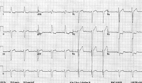
Left ventricular hypertrophy (LVH): Markedly increased LV voltages: huge precordial R and S waves that overlap with the adjacent leads (SV2 + RV6 >> 35 mm). R-wave peak time > 50 ms in V5-6 with associated QRS broadening. LV strain pattern with ST .
R Wave Peak Time Rwpt - Left Ventricular Hypertrophy (LVH) • LITFL • ECG .
ECG Pearl. There are no universally accepted criteria for diagnosing RVH in .ECG Criteria for Left Atrial Enlargement. LAE produces a broad, bifid P wave in .In LBBB, conduction delay means that impulses travel first via the right bundle .
U Waves - Left Ventricular Hypertrophy (LVH) • LITFL • ECG Library DiagnosisLeft Axis Deviation - Left Ventricular Hypertrophy (LVH) • LITFL • ECG .The most common causes of left ventricular hypertrophy are aortic stenosis, aortic regurgitation, hypertension, cardiomyopathy and coarctation of the aorta. .Electrocardiographic (ECG) left ventricular hypertrophy (LVH) with strain pattern is said to be present when, apart from the voltage criterion for ECG-LVH, there is also a downsloping .
what is lvh on ecg
Left ventricular hypertrophy (LVH): Markedly increased LV voltages: huge precordial R and S waves that overlap with the adjacent leads (SV2 + RV6 >> 35 mm). R-wave peak time > 50 ms in V5-6 with associated QRS broadening. LV strain pattern with ST depression and T-wave inversions in I, aVL and V5-6.The most common causes of left ventricular hypertrophy are aortic stenosis, aortic regurgitation, hypertension, cardiomyopathy and coarctation of the aorta. There are several ECG indexes, which generally have high diagnostic specificity but low sensitivity.Electrocardiographic (ECG) left ventricular hypertrophy (LVH) with strain pattern is said to be present when, apart from the voltage criterion for ECG-LVH, there is also a downsloping asymmetrical ST-segment depression with inverted asymmetric T wave ≥ 0.1 mV opposite the QRS axis in a resting ECG.
lvh with strain pattern meaning
LVH with strain pattern can sometimes be seen in long standing severe aortic regurgitation, usually with associated left ventricular hypertrophy and systolic dysfunction. The sensitivity of LVH strain pattern on ECG as a measure of LVH has ranged from 3.8% to 50% in various reports [1]. The electrocardiogram (ECG) is a useful but imperfect tool for detecting LVH. The utility of the ECG relates to its being relatively inexpensive and widely available. The limitations of the ECG relate to its moderate sensitivity or specificity depending upon which of the many proposed sets of diagnostic criteria are applied [ 1,2 ].
ECG strain was associated with increases in LV end‐diastolic volume, end‐systolic volume, mass, MVR (model 1, all P <0.05) and a decrease in LV EF (P <0.001) with highest standardized β‐coefficients for LV Δmass index (standardized β‐coefficient=0.084; Table 3). The ECG diagnosis of LVH is predominantly based on the QRS voltage criteria. The classical paradigm postulates that the increased left ventricular mass generates a stronger electrical field, increasing the leftward and posterior QRS forces, reflected in .
When LVH is caused by a pathological condition, we often see the "strain" pattern, which is ST depression and T wave inversion in leads with upright QRS complexes (the lateral leads). A reciprocal ST elevation can occur in the right-sided leads, which can be confusing to those looking for STEMI. In order to diagnose LVH from the ECG, we must also show repolarization abnormalities, called the "strain pattern". This is seen in sloping ST depressions in all leads with upright QRS complexes. There will also be slight ST elevations (reciprocal to the depressions) in leads with negative QRSs.By Steven Lome, MD. Enlarge. Left ventricular hypertrophy with strain pattern.
Left ventricular hypertrophy (LVH): Markedly increased LV voltages: huge precordial R and S waves that overlap with the adjacent leads (SV2 + RV6 >> 35 mm). R-wave peak time > 50 ms in V5-6 with associated QRS broadening. LV strain pattern with ST depression and T-wave inversions in I, aVL and V5-6.The most common causes of left ventricular hypertrophy are aortic stenosis, aortic regurgitation, hypertension, cardiomyopathy and coarctation of the aorta. There are several ECG indexes, which generally have high diagnostic specificity but low sensitivity.Electrocardiographic (ECG) left ventricular hypertrophy (LVH) with strain pattern is said to be present when, apart from the voltage criterion for ECG-LVH, there is also a downsloping asymmetrical ST-segment depression with inverted asymmetric T wave ≥ 0.1 mV opposite the QRS axis in a resting ECG.
LVH with strain pattern can sometimes be seen in long standing severe aortic regurgitation, usually with associated left ventricular hypertrophy and systolic dysfunction. The sensitivity of LVH strain pattern on ECG as a measure of LVH has ranged from 3.8% to 50% in various reports [1]. The electrocardiogram (ECG) is a useful but imperfect tool for detecting LVH. The utility of the ECG relates to its being relatively inexpensive and widely available. The limitations of the ECG relate to its moderate sensitivity or specificity depending upon which of the many proposed sets of diagnostic criteria are applied [ 1,2 ]. ECG strain was associated with increases in LV end‐diastolic volume, end‐systolic volume, mass, MVR (model 1, all P <0.05) and a decrease in LV EF (P <0.001) with highest standardized β‐coefficients for LV Δmass index (standardized β‐coefficient=0.084; Table 3).

premio sicuro givenchy crema viso
The ECG diagnosis of LVH is predominantly based on the QRS voltage criteria. The classical paradigm postulates that the increased left ventricular mass generates a stronger electrical field, increasing the leftward and posterior QRS forces, reflected in .
When LVH is caused by a pathological condition, we often see the "strain" pattern, which is ST depression and T wave inversion in leads with upright QRS complexes (the lateral leads). A reciprocal ST elevation can occur in the right-sided leads, which can be confusing to those looking for STEMI. In order to diagnose LVH from the ECG, we must also show repolarization abnormalities, called the "strain pattern". This is seen in sloping ST depressions in all leads with upright QRS complexes. There will also be slight ST elevations (reciprocal to the depressions) in leads with negative QRSs.
lvh voltage criteria by age
D is the fourth (number 4) letter in the alphabet. It comes from the Greek Delta and the Phoenician Dalet. Meanings for D. In education, D is a failing grade; In electronics, D is a standard size dry cell battery. In music, D is a note sometimes named “Re”. In Roman numerals, D also means the number 500.
lv strain pattern ecg|lvh strain pattern vs ischemia


